Health Guide

Tethered Spinal Cord Syndrome: Symptoms, Diagnosis, Treatment & How It Differs from PLID

26/09/2026

Dr. Saddam Hossain, PT

Medically reviewed by Dr. Saddam Hossain, PT

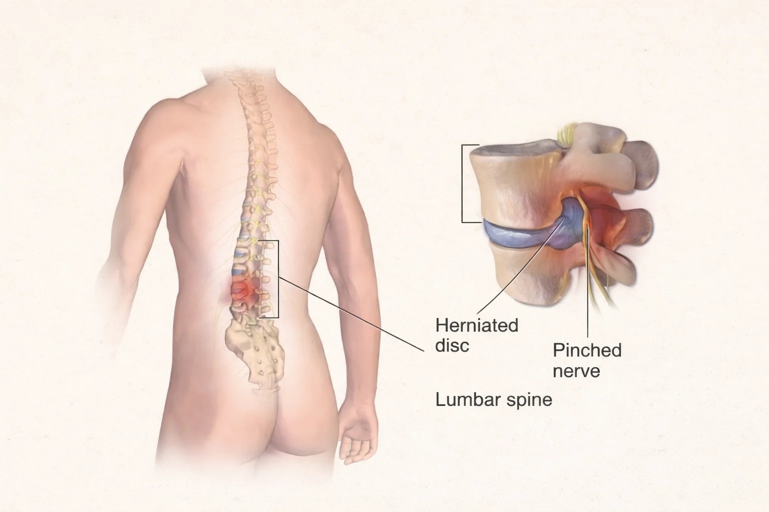
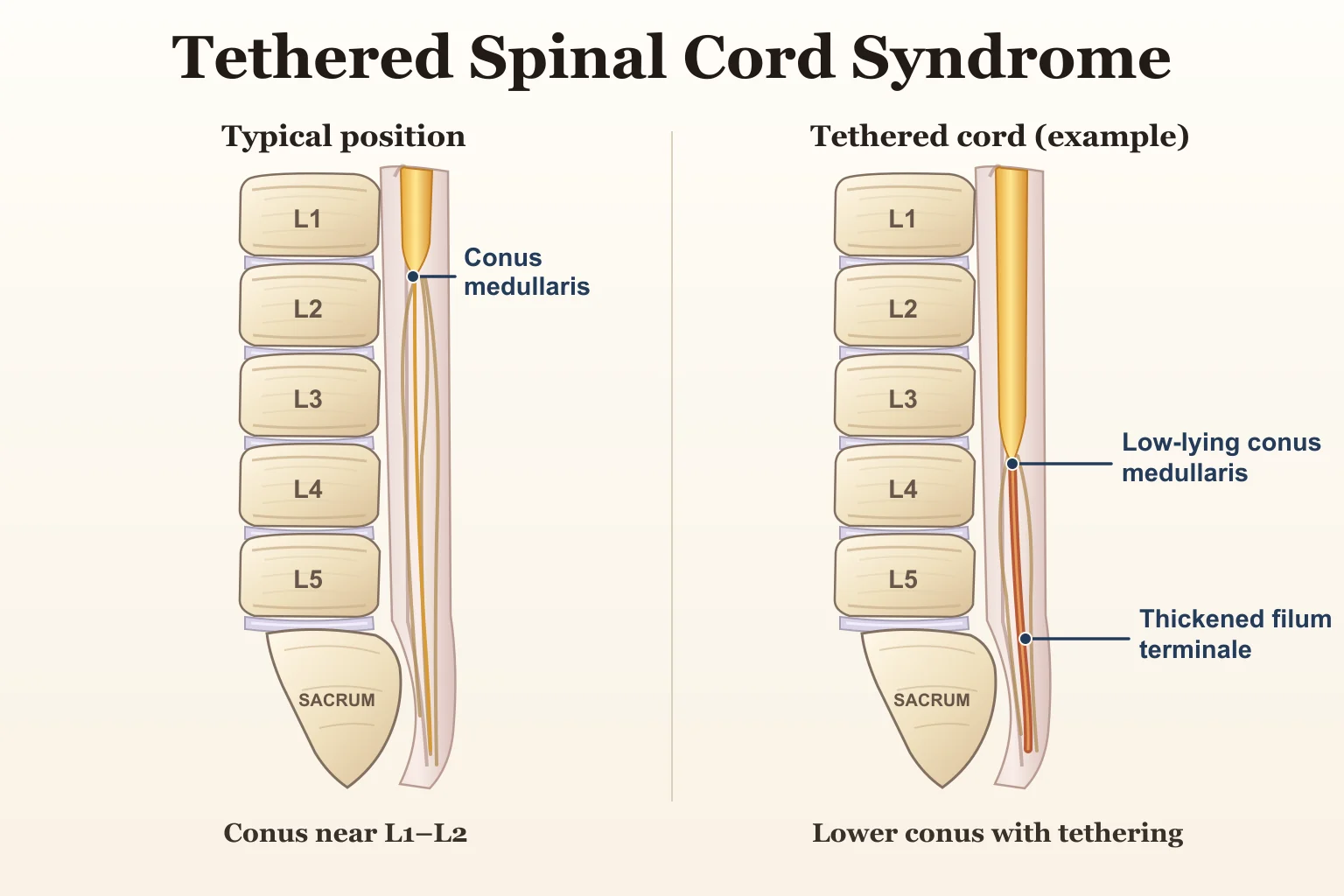
Schematic comparison showing five lumbar vertebrae and one fused sacrum, with a typical conus position and an example low-lying tethered cord

Low-back pain, leg pain, numbness, tingling, or weakness are commonly associated with conditions such as lumbar disc herniation or PLID. In many patients, these symptoms are indeed related to a disc problem. However, not every neurological symptom involving the lower back and legs originates from an intervertebral disc.

In some cases, symptoms may be related to a less common neurological condition known as Tethered Spinal Cord Syndrome (TCS). Because TCS can also cause back pain, leg symptoms, weakness, gait changes, and sensory disturbances, its presentation may sometimes resemble lumbar radiculopathy or PLID.

The distinction is clinically important. A disc abnormality seen on MRI does not automatically prove that it explains every symptom a patient is experiencing. When progressive weakness, changes in walking, foot deformity, bladder or bowel problems, or neurological findings that do not match the expected nerve-root pattern are present, a broader neurological assessment may be necessary. New bladder or bowel problems or saddle numbness with back or leg symptoms require emergency medical assessment.

This article explains what Tethered Spinal Cord Syndrome is, why it develops, how it affects the nervous system, how it is diagnosed and treated, and why differentiating it from PLID can sometimes be challenging.

What Is Tethered Spinal Cord Syndrome?

The spinal cord normally lies within the spinal canal with enough mobility to accommodate normal movement and growth of the spine. In Tethered Spinal Cord Syndrome, the spinal cord becomes abnormally fixed or attached to surrounding tissues, limiting this normal mobility.

As a result, abnormal longitudinal tension may develop within the spinal cord and its associated neural structures. Over time, this tension can interfere with neurological function and lead to symptoms involving movement, sensation, pain, walking ability, bladder and bowel control, and musculoskeletal alignment.

TCS can occur in children as part of congenital spinal abnormalities, but symptoms may also become apparent later in adolescence or adulthood.

Tethered Cord vs Tethered Cord Syndrome

These terms are related but are not always identical.

Tethered cord describes an anatomical situation in which the spinal cord is abnormally fixed or restricted.

Tethered Cord Syndrome refers to the clinical neurological dysfunction that develops when this tethering produces symptoms.

Therefore, an anatomical abnormality on imaging should not be considered in isolation. The patient's symptoms, neurological findings, functional changes, and imaging results must be interpreted together.

Understanding the Normal Spinal Cord and Conus Medullaris

To understand TCS, it is helpful to first understand several structures at the lower end of the spinal cord.

  1. Conus Medullaris:
    The conus medullaris is the tapered lower end of the spinal cord. In adults, it usually terminates around the upper lumbar spine, commonly near the L1-L2 level, although normal anatomical variation exists.
  2. Filum Terminale:
    The filum terminale is a thin fibrous structure extending downward from the conus medullaris. It helps stabilize the spinal cord within the spinal canal.
  3. Cauda Equina:
    Below the end of the spinal cord, the lumbar and sacral nerve roots continue downward as a bundle known as the cauda equina.

Normally, these structures have sufficient freedom to accommodate spinal movement. In tethering disorders, abnormal fixation may reduce this mobility and place excessive tension on neural tissues.

What Causes Tethered Spinal Cord Syndrome?

Tethering may be related to congenital abnormalities or may develop secondarily after surgery or other spinal pathology.

Congenital Causes

Congenital causes include abnormalities that develop during formation of the spinal cord and surrounding structures. These may include:

  1. Thickened filum terminale:
    The filum becomes abnormally thick and may restrict normal spinal cord movement.
  2. Fatty filum terminale:
    Fatty tissue within the filum may be associated with abnormal tethering.
  3. Spinal lipoma:
    A fatty mass may attach to or distort neural structures and contribute to tethering.
  4. Lipomyelomeningocele:
    A congenital spinal dysraphism in which fatty tissue is connected to the spinal cord through a defect in the posterior spinal elements.
  5. Myelomeningocele:
    A severe form of spinal dysraphism in which neural tissue and meninges protrude through a spinal defect.
  6. Split cord malformation:
    The spinal cord may be divided into two parts by an abnormal septum or bony/cartilaginous structure.
  7. Other spinal dysraphisms:
    Various congenital abnormalities involving incomplete or abnormal development of the spine and spinal cord may lead to tethering.

Acquired or Secondary Tethering

Tethering can also occur later in life due to:

  1. Previous spinal surgery
  2. Postoperative scar tissue
  3. Adhesions around neural structures
  4. Spinal trauma
  5. Retethering after previous untethering surgery

Postoperative retethering is particularly relevant in patients who previously underwent surgery for congenital spinal dysraphism.

How Does Tethering Damage the Spinal Cord?

The basic mechanism can be understood as:

Abnormal fixation of the spinal cord → increased longitudinal tension → stretching of neural tissue and its microcirculation → progressive neurological dysfunction

When the spinal cord is abnormally fixed, normal movement or spinal growth may increase traction on the neural tissues. This can interfere with neural function and, over time, contribute to weakness, sensory disturbance, pain, gait abnormalities, or bladder and bowel dysfunction.

In children, symptoms may become more noticeable during periods of growth because the vertebral column lengthens while the tethered spinal cord cannot adapt normally.

Symptoms of Tethered Cord Syndrome

The presentation of TCS varies considerably depending on age, underlying anatomy, severity of tethering, and the neurological structures involved.

Some patients mainly experience pain, while others present with progressive weakness, urinary dysfunction, gait changes, or orthopedic deformity.

Neurological Symptoms

Neurological manifestations may include:

  1. Lower-limb weakness
  2. Progressive weakness over time
  3. Muscle wasting
  4. Numbness or altered sensation
  5. Tingling or paresthesia
  6. Abnormal reflexes
  7. Changes in muscle control
  8. Progressive neurological deficit

The pattern may be asymmetric and does not always correspond neatly to a single lumbar nerve root.

Pain

Pain may occur in:

  1. The lower back
  2. One or both legs
  3. The sacral region
  4. The buttock or perineal region in selected cases

Unlike classic radiculopathy, the pain pattern may sometimes be difficult to assign to a single dermatome or nerve root.

Gait and Mobility Changes

TCS may affect walking and functional mobility.

Patients may develop:

  1. Altered gait pattern
  2. Difficulty walking
  3. Reduced walking endurance
  4. Frequent falls
  5. Progressive foot drop or lower-limb weakness
  6. Increasing difficulty with daily mobility

A progressive change in gait is particularly important because it may indicate worsening neurological function.

Bladder and Bowel Symptoms

Bladder dysfunction is one of the clinically important manifestations of TCS.

Possible symptoms include:

  1. Urinary urgency
  2. Increased urinary frequency
  3. Urinary incontinence
  4. Difficulty initiating urination
  5. Urinary retention
  6. Incomplete bladder emptying
  7. Recurrent urinary problems or infections
  8. Bowel dysfunction

When the sacral neural pathways responsible for bladder control are affected, a neurogenic bladder may develop. This means that the bladder does not store or empty urine normally because of neurological dysfunction rather than a primary bladder disorder.

New bladder or bowel problems with back or leg symptoms, especially urinary retention or saddle numbness, require emergency medical assessment to exclude cauda equina syndrome and other serious causes.

Orthopedic Findings

Longstanding neurological dysfunction may also produce musculoskeletal changes, including:

  1. Scoliosis
  2. Pes cavus
  3. Clubfoot
  4. Contractures
  5. Asymmetry of the lower limbs
  6. Progressive foot deformity
  7. Abnormal posture or limb alignment

These findings can be especially important in children and adolescents.

Signs of Tethered Cord Syndrome in Children

In children, TCS may not initially present as a typical back-pain condition.

Possible signs include:

  1. Abnormal gait
  2. Delayed development of walking or motor skills
  3. Progressive foot deformity
  4. Scoliosis
  5. Lower-limb weakness
  6. Loss of previously established bladder control
  7. Recurrent urinary problems
  8. Lumbosacral skin abnormalities

A child whose walking pattern, lower-limb strength, foot alignment, or bladder function progressively changes should receive appropriate neurological evaluation rather than being managed only as a local orthopedic problem.

Skin Signs That May Suggest Occult Spinal Dysraphism

Some congenital spinal abnormalities may be associated with visible skin findings over the lumbosacral region.

These may include:

  1. A tuft of hair
  2. An atypical or deep sacral dimple
  3. A subcutaneous fatty lump
  4. Hemangioma
  5. Abnormal skin pigmentation
  6. Dermal sinus
  7. Other unusual skin abnormalities over the lower spine

These findings may suggest an underlying spinal dysraphism and warrant further medical assessment when clinically appropriate.

However, an important point is that the absence of visible skin abnormalities does not exclude Tethered Cord Syndrome.

Why Can Tethered Cord Syndrome Be Confused with PLID?

TCS and PLID are fundamentally different disorders, but some of their symptoms overlap.

Both conditions may present with:

  1. Low-back pain
  2. Leg pain
  3. Numbness
  4. Tingling
  5. Lower-limb weakness
  6. Difficulty walking
  7. Neurological symptoms

This overlap can make the initial clinical picture appear similar.

The diagnostic challenge becomes greater when lumbar MRI shows a disc bulge or herniation. Disc abnormalities are common, especially with increasing age, and not every imaging finding is necessarily responsible for every symptom.

The key question is therefore not simply:

“Is there a disc abnormality?”

The more useful question is:

“Does this abnormality adequately explain the patient's neurological pattern?”

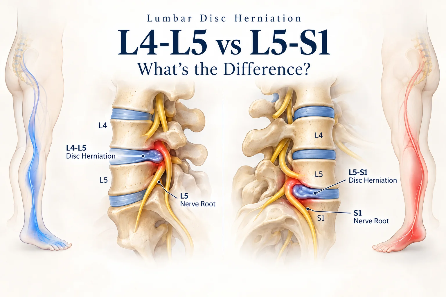
For example, if an MRI shows an L4-L5 disc lesion but the patient has bilateral progressive weakness, bladder dysfunction, an abnormal gait, longstanding foot deformity, or sensory findings that do not correspond to the expected nerve-root distribution, the disc lesion may not explain the entire clinical picture.

That is when alternative neurological causes should be considered.

Tethered Cord Syndrome vs PLID

Although both conditions may produce back and leg symptoms, their underlying pathology and neurological patterns are different.

FeatureTethered Cord SyndromePLID / Lumbar Disc Herniation
Primary pathologyAbnormal fixation and traction of the spinal cordDisplacement or herniation of an intervertebral disc affecting nearby neural structures
Pain patternMay be diffuse, atypical, bilateral, or non-dermatomalOften follows a specific nerve-root distribution
WeaknessMay involve multiple muscles or neurological levelsMore often follows the affected nerve root or myotome
Sensory disturbanceVariable and may not follow a single dermatomeOften corresponds to a dermatome
Bladder dysfunctionImportant possible manifestationUnusual in uncomplicated PLID; requires urgent concern if cauda equina syndrome is suspected
Gait deteriorationMay progressively developMay occur with significant radiculopathy or motor deficit
Foot deformityMay occur, particularly with longstanding congenital diseaseNot a typical feature
ScoliosisMay be associatedNot a characteristic diagnostic feature
Lumbosacral skin markersMay be presentNot associated with PLID
MRI findingsMay show low conus, abnormal filum, lipoma, split cord or other dysraphismMay show disc protrusion, extrusion or sequestration
Treatment approachNeurological/neurosurgical assessment; untethering when indicatedUsually conservative initially, with surgery reserved for selected cases

For a fuller explanation of disc prolapse, see our PLID pillar guide.

When “PLID” May Not Explain the Whole Clinical Picture

A diagnosis of PLID should be questioned or reassessed when the clinical findings do not correlate well with the expected disc-related pattern.

Features that may warrant further evaluation include:

  1. Progressive bilateral weakness:
    A single lumbar nerve-root lesion may not adequately explain progressive involvement of both lower limbs.
  2. Symptoms involving multiple neurological levels:
    Weakness or sensory disturbance affecting several myotomes or dermatomes should prompt broader neurological consideration.
  3. Neurological findings that do not match the MRI level:
    Imaging and clinical examination should make anatomical sense together.
  4. Progressive gait deterioration:
    A worsening walking pattern can indicate neurological progression.
  5. Foot deformity:
    Longstanding or progressive foot deformity is not typical of a simple acute lumbar disc prolapse.
  6. Bladder or bowel changes:
    These symptoms require careful neurological assessment.
  7. Childhood spinal abnormalities:
    Previous spinal dysraphism, congenital deformity, surgery, or unusual skin markers may provide an important clue.
  8. Failure to improve as expected:
    Persistent or worsening neurological symptoms despite appropriate management should trigger reassessment.
  9. Atypical or non-dermatomal symptoms:
    Symptoms that do not follow one recognizable nerve-root pattern may indicate a different neurological process.

The central principle is:

Clinical findings should correlate with imaging findings.

An MRI should support the clinical diagnosis rather than replace clinical reasoning.

Can Tethered Cord and PLID Exist Together?

Yes. These conditions are not necessarily mutually exclusive.

A patient may have:

Tethered spinal cord + lumbar disc degeneration or disc herniation.

This creates a more complex diagnostic situation.

For example, a disc herniation may explain a particular radicular pain pattern, while progressive bladder dysfunction, bilateral neurological changes, or longstanding foot deformity may originate from an additional neurological condition.

Therefore, the diagnostic question may not always be:

“Is this TCS or PLID?”

Sometimes the more appropriate question is:

Which abnormality is responsible for which symptoms?

This is why neurological examination and clinical-radiological correlation remain essential.

How Is Tethered Cord Syndrome Diagnosed?

TCS is diagnosed through a combination of clinical assessment, neurological examination, imaging, and selected additional investigations.

Clinical Assessment

A detailed assessment may include:

  1. History and progression of symptoms
  2. Lower-limb muscle power
  3. Sensory examination
  4. Deep tendon reflexes
  5. Muscle tone
  6. Gait analysis
  7. Foot and lower-limb alignment
  8. Spinal deformity assessment
  9. Bladder and bowel history
  10. Lumbosacral skin examination
  11. Previous congenital spinal conditions
  12. History of spinal surgery

The pattern and progression of symptoms are often just as important as their presence.

MRI

MRI is the principal imaging investigation used to evaluate suspected tethering.

Possible MRI findings may include:

  1. Low-lying conus medullaris
  2. Thickened filum terminale
  3. Fat-containing filum
  4. Spinal lipoma
  5. Lipomyelomeningocele
  6. Split cord malformation
  7. Other associated spinal dysraphism

However, TCS should not be diagnosed solely on the basis of one imaging feature. Imaging must be interpreted together with neurological and functional findings.

Urodynamic Assessment

Urodynamic testing may be recommended when bladder dysfunction is suspected.

It can help assess:

  • bladder storage function
  • bladder pressure
  • detrusor activity
  • urinary emptying
  • neurogenic bladder dysfunction

This can be particularly important in children and in patients with longstanding urinary symptoms.

Other Investigations

Depending on the clinical presentation, additional investigations may include:

  1. Spinal ultrasound in young infants
  2. Renal or urinary tract evaluation
  3. Electrodiagnostic testing in selected cases
  4. X-ray for scoliosis or skeletal deformity
  5. Other specialist neurological or urological investigations

Conditions That Can Mimic Tethered Cord Syndrome

TCS is not the only condition capable of causing back pain, lower-limb symptoms, or neurological dysfunction.

Differential diagnoses may include:

  1. Lumbar disc herniation / PLID:
    May produce nerve-root pain, weakness, and sensory changes.
  2. Lumbar spinal stenosis:
    Can cause leg pain, weakness, and walking limitation, particularly in older adults.
  3. Cauda equina syndrome:
    A neurological emergency involving severe compression of the cauda equina, often associated with bladder/bowel dysfunction and saddle sensory changes.
  4. Peripheral neuropathy:
    May cause weakness, numbness, or sensory disturbance in the lower limbs.
  5. Other spinal cord disorders:
    Structural, inflammatory, congenital, or compressive neurological disorders can sometimes produce similar findings.
  6. Musculoskeletal low-back pain:
    Mechanical back pain is much more common but generally does not explain progressive neurological, bladder, or bowel dysfunction.

Treatment of Tethered Cord Syndrome

Treatment depends on the patient's age, symptoms, underlying anatomy, neurological progression, bladder function, and specialist assessment.

Not every anatomical tethering abnormality automatically requires surgery.

Observation and Monitoring

Selected patients may be monitored when symptoms are absent, minimal, or stable.

Monitoring may involve:

  • neurological examination
  • assessment of gait and strength
  • bladder evaluation
  • orthopedic monitoring
  • repeat imaging or specialist follow-up when indicated

Surgical Untethering

For symptomatic or progressive TCS, surgical untethering may be considered.

During the procedure, the neurosurgeon releases the structures that are placing abnormal tension on the spinal cord.

The goals of surgery may include:

  1. Reducing pathological spinal cord tension
  2. Preventing further neurological deterioration
  3. Preserving motor function
  4. Preserving bladder and bowel function
  5. Reducing pain in selected patients
  6. Limiting progression of deformity or functional loss

The aim is not always to completely reverse every established neurological deficit. Longstanding weakness, sensory loss, or bladder dysfunction may persist even after successful surgery.

Role of Physiotherapy in Tethered Cord Syndrome

A crucial point for patients and clinicians is:

Physiotherapy does not release or “untether” the spinal cord.

Physiotherapy is therefore not a substitute for neurological or neurosurgical assessment when symptomatic tethering is suspected.

Its role is primarily in rehabilitation, preservation of function, management of secondary impairments, and postoperative recovery.

Depending on the patient's neurological status, physiotherapy may include:

  1. Strengthening preserved musculature:
    Exercises may target lower-limb and trunk muscles according to the patient's strength and neurological status.
  2. Maintaining joint mobility:
    Appropriate range-of-motion exercises can help prevent stiffness.
  3. Contracture prevention:
    Positioning, stretching, mobility work, and orthotic management may be used when indicated.
  4. Balance rehabilitation:
    Static and dynamic balance training can improve safety and mobility.
  5. Gait training:
    Patients with weakness or altered gait may benefit from task-specific walking rehabilitation.
  6. Assistive-device training:
    Canes, crutches, walkers, or other mobility aids may be introduced when appropriate.
  7. Orthotic management:
    Foot orthoses, AFOs, or other devices may help improve alignment and walking efficiency in selected patients.
  8. Functional rehabilitation:
    Treatment should focus on meaningful activities such as transfers, walking, stairs, self-care, school, work, or community mobility.
  9. Postoperative rehabilitation:
    Following neurosurgical clearance, physiotherapy may assist with gradual restoration of strength, mobility, balance, and function.

The rehabilitation plan should be individualized according to the patient's neurological findings, functional limitations, medical status, and treatment goals.

Physiotherapy Precautions in Suspected Tethered Cord Syndrome

Patients with suspected or confirmed TCS should not automatically be managed as routine mechanical low-back pain.

Particular caution is appropriate with:

  1. Aggressive spinal traction
  2. Forceful spinal stretching
  3. High-velocity spinal manipulation
  4. Exercises that significantly worsen neurological symptoms
  5. Repeated routine back-pain treatment despite progressive neurological deterioration

Most importantly, new bladder or bowel dysfunction, saddle numbness, or rapidly worsening leg weakness require emergency medical assessment; other progressive neurological changes need prompt specialist review rather than more routine back-pain treatment.

Red Flags That Need Further Medical Evaluation

Seek timely medical or neurological evaluation if back or leg symptoms are associated with:

  1. Progressive lower-limb weakness
  2. New urinary retention or incontinence
  3. New bowel dysfunction
  4. Rapid deterioration in walking
  5. Progressive foot deformity
  6. Significant or worsening sensory loss
  7. Increasing scoliosis
  8. Neurological findings that do not match the presumed disc lesion

These findings do not automatically confirm TCS. New urinary retention, loss of bladder or bowel control, or saddle numbness with back or leg symptoms requires emergency assessment because cauda equina syndrome is possible. Other progressive neurological signs warrant prompt specialist assessment.

What Should a Physiotherapist Assess?

Physiotherapists often encounter patients with low-back pain, leg pain, weakness, gait problems, and mobility limitations. A structured assessment can help determine whether the presentation appears mechanical or whether further medical investigation is necessary.

Assessment may include:

  1. Detailed symptom history
  2. Progression of neurological symptoms
  3. Muscle power
  4. Sensation
  5. Reflexes
  6. Gait pattern
  7. Balance
  8. Foot alignment and deformity
  9. Spinal alignment
  10. Functional limitations
  11. Bladder and bowel red-flag screening
  12. Correlation between clinical findings and available imaging

The physiotherapist's role is not to independently confirm TCS from symptoms alone, but to recognize when a presentation does not behave like uncomplicated musculoskeletal low-back pain and refer appropriately.

Can Tethered Cord Syndrome Be Completely Cured?

The outcome of TCS varies considerably between patients.

Factors influencing recovery may include:

  1. The underlying cause of tethering
  2. Age at diagnosis
  3. Duration of symptoms
  4. Severity of neurological impairment
  5. Degree of bladder involvement
  6. Presence of musculoskeletal deformity
  7. Timing of surgery when surgery is indicated
  8. Risk of retethering

Some patients experience meaningful improvement after appropriate treatment, while others may continue to have residual weakness, sensory changes, bladder dysfunction, or orthopedic problems.

For this reason, it is more accurate to discuss neurological preservation, functional improvement, and prevention of further deterioration rather than promising a complete cure.

Retethering After Surgery

Even after successful untethering, the spinal cord may become tethered again.

This is known as retethering.

Scar tissue and postoperative adhesions may gradually restrict neural movement again, particularly in patients with complex congenital spinal dysraphism.

Possible signs of retethering may include:

  • renewed pain
  • progressive weakness
  • worsening gait
  • new sensory changes
  • bladder deterioration
  • progressive orthopedic deformity

Long-term follow-up may therefore be necessary in selected patients.

When Should You Seek Professional Evaluation?

Persistent low-back pain alone is common and is usually not caused by TCS. However, professional evaluation becomes particularly important when back or leg symptoms occur together with neurological or urological changes.

Seek further assessment if you notice:

  1. Progressive weakness in one or both legs
  2. New or worsening walking difficulty
  3. Recurrent falls or foot drop
  4. Progressive foot deformity
  5. New urinary or bowel problems
  6. Unusual sensory loss or numbness
  7. Symptoms that do not follow a typical nerve-root pattern
  8. Neurological symptoms that do not correlate with the reported lumbar disc abnormality

New bladder or bowel dysfunction, saddle numbness, or rapidly worsening leg weakness requires emergency medical assessment. Other progressive neurological findings need prompt neurological or neurosurgical referral rather than continued routine back-pain treatment.

Conclusion

Tethered Spinal Cord Syndrome and PLID are very different conditions, but some of their symptoms can overlap. Both may cause low-back pain, leg pain, weakness, numbness, and walking difficulties, which is why careful clinical assessment is important.

In TCS, the fundamental problem is abnormal fixation and traction of the spinal cord, whereas in PLID the primary issue is intervertebral disc pathology affecting nearby neural structures.

The most important clinical lesson is that diagnosis should not depend on imaging alone. MRI findings must be interpreted alongside the patient's neurological examination, gait, sensory pattern, muscle weakness, bladder and bowel function, orthopedic findings, and symptom progression.

A disc abnormality on MRI should not automatically be assumed to explain every case of back pain, leg symptoms, or neurological dysfunction.

Why Asia Digital Physiotherapy & Orthopedic Rehabilitation Center (ADPORC) Is a Trusted Physiotherapy Center in Dhaka

ADPORC focuses on evidence-based physiotherapy for musculoskeletal, neurological, and sports-related conditions.

  1. Expert Care by Dr. Saddam Hossain, PT:
    Structured rehabilitation under experienced clinical supervision.
  2. Condition-Specific Physiotherapy Programs:
    Treatment plans are designed according to clinical assessment, pain stage, functional limitations, and rehabilitation goals.
  3. Advanced Electrotherapy and Rehabilitation Equipment:
    Modalities are used only when clinically indicated.
  4. One-on-One Supervised Sessions:
    Direct supervision helps ensure safe and appropriate rehabilitation.
  5. Accessible Location in Jatrabari, Dhaka:
    ADPORC serves patients from Jatrabari, Dholaipar, Shonir Akhra, Doyagonj, Puran Dhaka, Keraniganj, Narayanganj, and surrounding areas.

For patients with back pain, leg weakness, gait abnormalities, or neurological symptoms, assessment should focus first on identifying the underlying cause and determining whether physiotherapy, medical referral, or multidisciplinary management is most appropriate.

Book Your Appointment Today

Do not ignore persistent back pain or progressive neurological symptoms. Early assessment can help identify whether the problem is mechanical, neurological, or requires further specialist evaluation.

Visit ADPORC for professional physiotherapy assessment and rehabilitation planning.

কোমর ব্যথা, পায়ে ব্যথা, ঝিনঝিনি, অবশভাব বা দুর্বলতা দেখা দিলে অনেক সময় PLID বা লাম্বার ডিস্ক হার্নিয়েশনকে সম্ভাব্য কারণ হিসেবে ধরা হয়। অনেক রোগীর ক্ষেত্রে সত্যিই ডিস্ক-সংক্রান্ত সমস্যাই এসব উপসর্গের কারণ হয়। তবে কোমর ও পায়ের প্রতিটি স্নায়বিক উপসর্গ যে ডিস্কের সমস্যার কারণেই হবে, তা নয়।

কিছু ক্ষেত্রে এসব উপসর্গের পেছনে থাকতে পারে তুলনামূলকভাবে কম পরিচিত একটি স্নায়বিক সমস্যা, যার নাম টেথার্ড স্পাইনাল কর্ড সিনড্রোম (Tethered Spinal Cord Syndrome বা TCS)। এই অবস্থায়ও কোমর ব্যথা, পায়ে ব্যথা, দুর্বলতা, হাঁটার ধরনে পরিবর্তন এবং অনুভূতির সমস্যা দেখা দিতে পারে। ফলে কখনও কখনও এটি PLID বা লাম্বার র‍্যাডিকুলোপ্যাথির মতো মনে হতে পারে।

এখানে গুরুত্বপূর্ণ বিষয় হলো, MRI-তে ডিস্ক বাল্জ বা ডিস্ক হার্নিয়েশন দেখা গেলেই সেটিকেই রোগীর সব উপসর্গের একমাত্র কারণ ধরে নেওয়া উচিত নয়। বিশেষ করে যদি ধীরে ধীরে পায়ের দুর্বলতা বাড়ে, হাঁটার ধরন বদলে যায়, পায়ের গঠনে বিকৃতি দেখা দেয়, প্রস্রাব-পায়খানার নিয়ন্ত্রণে সমস্যা হয়, অথবা স্নায়বিক পরীক্ষার ফলাফল সম্ভাব্য নার্ভ রুটের সঙ্গে না মেলে, তাহলে আরও বিস্তারিত স্নায়বিক মূল্যায়ন প্রয়োজন হতে পারে। কোমর বা পায়ের উপসর্গের সঙ্গে নতুন প্রস্রাব-পায়খানার সমস্যা বা স্যাডল অঞ্চলে অবশভাব দেখা দিলে জরুরি চিকিৎসা মূল্যায়ন নিন।

এই ব্লগে আমরা আলোচনা করব টেথার্ড স্পাইনাল কর্ড সিনড্রোম কী, কেন হয়, কী কী লক্ষণ দেখা দিতে পারে, কীভাবে রোগ নির্ণয় করা হয়, কী ধরনের চিকিৎসা প্রয়োজন হতে পারে এবং PLID থেকে এটি কীভাবে আলাদা।

টেথার্ড স্পাইনাল কর্ড সিনড্রোম কী?

স্বাভাবিক অবস্থায় স্পাইনাল কর্ড স্পাইনাল ক্যানালের ভেতরে এমনভাবে অবস্থান করে, যাতে শরীরের বৃদ্ধি ও মেরুদণ্ডের স্বাভাবিক নড়াচড়ার সঙ্গে এটি কিছুটা মানিয়ে নিতে পারে।

টেথার্ড স্পাইনাল কর্ড সিনড্রোমে স্পাইনাল কর্ড আশপাশের কোনো টিস্যুর সঙ্গে অস্বাভাবিকভাবে আটকে যায় বা স্থির হয়ে যায়। ফলে স্পাইনাল কর্ডের স্বাভাবিক নড়াচড়া কমে যায় এবং এতে অস্বাভাবিক টান তৈরি হতে পারে।

সময়ের সঙ্গে এই টান স্নায়ুতন্ত্রের স্বাভাবিক কার্যকারিতায় বাধা সৃষ্টি করতে পারে। এর ফলে চলাফেরা, অনুভূতি, ব্যথা, হাঁটার ক্ষমতা, প্রস্রাব-পায়খানার নিয়ন্ত্রণ এবং শরীরের অ্যালাইনমেন্টে সমস্যা দেখা দিতে পারে।

TCS শিশুদের মধ্যে জন্মগত স্পাইনাল সমস্যার সঙ্গে দেখা যেতে পারে, আবার কৈশোর বা প্রাপ্তবয়স্ক বয়সেও এর উপসর্গ প্রথমবার স্পষ্ট হতে পারে।

টেথার্ড কর্ড ও টেথার্ড কর্ড সিনড্রোম কি একই?

দুটি শব্দ সম্পর্কিত হলেও একেবারে একই বিষয় বোঝায় না।

টেথার্ড কর্ড বলতে বোঝায় এমন একটি শারীরবৃত্তীয় অবস্থা, যেখানে স্পাইনাল কর্ড অস্বাভাবিকভাবে আটকে বা সীমাবদ্ধ হয়ে যায়।

অন্যদিকে টেথার্ড কর্ড সিনড্রোম বলতে বোঝায় সেই ক্লিনিক্যাল স্নায়বিক সমস্যা, যা এই টেথারিংয়ের কারণে উপসর্গ তৈরি হলে দেখা দেয়।

তাই শুধু MRI-তে অস্বাভাবিকতা দেখা গেলেই রোগ নির্ণয় করা উচিত নয়। রোগীর উপসর্গ, স্নায়বিক পরীক্ষা, দৈনন্দিন কাজের সীমাবদ্ধতা এবং ইমেজিংয়ের ফলাফল একসঙ্গে বিবেচনা করতে হয়।

স্পাইনাল কর্ড, কনাস মেডুলারিস ও ফিলাম টার্মিনালে

TCS বুঝতে হলে স্পাইনাল কর্ডের নিচের অংশের কয়েকটি গুরুত্বপূর্ণ গঠন সম্পর্কে ধারণা থাকা দরকার।

  1. কনাস মেডুলারিস:
    কনাস মেডুলারিস হলো স্পাইনাল কর্ডের নিচের দিকে সরু হয়ে যাওয়া শেষ অংশ। প্রাপ্তবয়স্কদের ক্ষেত্রে এটি সাধারণত L1-L2 স্তরের কাছাকাছি শেষ হয়, যদিও স্বাভাবিকভাবে কিছু ভিন্নতা থাকতে পারে।
  2. ফিলাম টার্মিনালে:
    ফিলাম টার্মিনালে হলো কনাস মেডুলারিস থেকে নিচের দিকে প্রসারিত একটি পাতলা তন্তুযুক্ত গঠন, যা স্পাইনাল কর্ডকে স্থিতিশীল রাখতে সাহায্য করে।
  3. কাউডা ইকুইনা:
    স্পাইনাল কর্ড শেষ হওয়ার পর লাম্বার ও স্যাক্রাল নার্ভ রুটগুলো নিচের দিকে একটি গুচ্ছের মতো নেমে যায়। এই অংশকে কাউডা ইকুইনা বলা হয়।

স্বাভাবিক অবস্থায় এসব গঠন মেরুদণ্ডের নড়াচড়ার সঙ্গে কিছুটা মানিয়ে নিতে পারে। কিন্তু টেথারিং থাকলে অস্বাভাবিক আটকে যাওয়ার কারণে স্পাইনাল কর্ডের ওপর অতিরিক্ত টান পড়তে পারে।

টেথার্ড স্পাইনাল কর্ড সিনড্রোম কেন হয়?

TCS জন্মগত কারণে হতে পারে, আবার পরবর্তীতে স্পাইনাল সার্জারি, স্কার টিস্যু বা অন্যান্য সমস্যার পরও তৈরি হতে পারে।

জন্মগত কারণ

  1. থিকেন্ড ফিলাম টার্মিনালে:
    ফিলাম অস্বাভাবিকভাবে মোটা হয়ে স্পাইনাল কর্ডের স্বাভাবিক নড়াচড়া কমিয়ে দিতে পারে।
  2. ফ্যাটি ফিলাম টার্মিনালে:
    ফিলামের মধ্যে অতিরিক্ত চর্বিযুক্ত টিস্যু টেথারিংয়ের সঙ্গে সম্পর্কিত হতে পারে।
  3. স্পাইনাল লাইপোমা:
    চর্বিযুক্ত একটি গঠন স্নায়বিক টিস্যুর সঙ্গে যুক্ত হয়ে টান সৃষ্টি করতে পারে।
  4. লাইপোমায়েলোমেনিনগোসিল:
    জন্মগত স্পাইনাল ডাইসরাফিজমের একটি ধরন, যেখানে চর্বিযুক্ত টিস্যু স্পাইনাল কর্ডের সঙ্গে যুক্ত থাকে।
  5. মায়েলোমেনিনগোসিল:
    গুরুতর ধরনের জন্মগত স্পাইনাল ডাইসরাফিজম।
  6. স্প্লিট কর্ড ম্যালফরমেশন:
    স্পাইনাল কর্ড অস্বাভাবিকভাবে দুই ভাগে বিভক্ত হতে পারে।
  7. অন্যান্য স্পাইনাল ডাইসরাফিজম:
    মেরুদণ্ড ও স্পাইনাল কর্ডের বিকাশজনিত বিভিন্ন অস্বাভাবিকতা টেথারিংয়ের কারণ হতে পারে।

পরবর্তী জীবনে তৈরি হওয়া কারণ

  1. আগের স্পাইনাল সার্জারি
  2. অপারেশনের পর স্কার টিস্যু
  3. স্নায়বিক গঠনের চারপাশে আঁটসাঁট জোড়া লেগে যাওয়া
  4. স্পাইনাল ট্রমা
  5. আগের আনটেথারিং সার্জারির পর পুনরায় টেথারিং

জন্মগত স্পাইনাল সমস্যার জন্য আগে সার্জারি করা রোগীদের মধ্যে পরবর্তীতে আবার টেথারিং হওয়া গুরুত্বপূর্ণ একটি বিষয়।

টেথারিং কীভাবে স্পাইনাল কর্ডের ক্ষতি করে?

এই সমস্যার মূল প্রক্রিয়া সহজভাবে এভাবে বোঝানো যায়:

স্পাইনাল কর্ড অস্বাভাবিকভাবে আটকে যাওয়া → দৈর্ঘ্য বরাবর টান বৃদ্ধি → স্নায়বিক টিস্যু ও ক্ষুদ্র রক্তনালির ওপর চাপ → স্নায়বিক কার্যকারিতা ধীরে ধীরে কমে যাওয়া

স্পাইনাল কর্ড যদি আটকে থাকে, তাহলে মেরুদণ্ডের নড়াচড়া বা শরীরের বৃদ্ধির সঙ্গে স্নায়বিক টিস্যুর ওপর টান বাড়তে পারে। এর ফলে সময়ের সঙ্গে দুর্বলতা, অনুভূতির সমস্যা, ব্যথা, হাঁটার পরিবর্তন এবং প্রস্রাব-পায়খানার নিয়ন্ত্রণের সমস্যা তৈরি হতে পারে।

শিশুদের ক্ষেত্রে বয়স বাড়ার সঙ্গে মেরুদণ্ডের দৈর্ঘ্য বাড়ে। কিন্তু টেথার্ড স্পাইনাল কর্ড স্বাভাবিকভাবে মানিয়ে নিতে না পারলে উপসর্গ আরও স্পষ্ট হয়ে উঠতে পারে।

টেথার্ড কর্ড সিনড্রোমের লক্ষণ

TCS-এর লক্ষণ রোগীর বয়স, টেথারিংয়ের ধরন ও তীব্রতা এবং কোন স্নায়বিক অংশ বেশি আক্রান্ত হয়েছে তার ওপর নির্ভর করে।

কিছু রোগীর প্রধান সমস্যা ব্যথা, আবার কারও ক্ষেত্রে দুর্বলতা, প্রস্রাবের সমস্যা, হাঁটার পরিবর্তন বা পায়ের গঠনে বিকৃতি বেশি স্পষ্ট হতে পারে।

স্নায়বিক লক্ষণ

  1. এক বা দুই পায়ে দুর্বলতা
  2. সময়ের সঙ্গে দুর্বলতা বাড়া
  3. পেশি শুকিয়ে যাওয়া
  4. অবশভাব বা অনুভূতি কমে যাওয়া
  5. ঝিনঝিনি অনুভূতি
  6. রিফ্লেক্সের পরিবর্তন
  7. পেশি নিয়ন্ত্রণে সমস্যা
  8. ধীরে ধীরে স্নায়বিক কার্যকারিতা কমে যাওয়া

সব সময় এসব লক্ষণ একটি নির্দিষ্ট নার্ভ রুটের সঙ্গে পুরোপুরি মেলে না।

ব্যথা

ব্যথা হতে পারে:

  1. কোমরের নিচের অংশে
  2. এক বা দুই পায়ে
  3. স্যাক্রাল অঞ্চলে
  4. কিছু ক্ষেত্রে নিতম্ব বা পেরিনিয়াল অঞ্চলে

ক্লাসিক র‍্যাডিকুলোপ্যাথির মতো সব সময় ব্যথা একটি নির্দিষ্ট ডার্মাটোম বা নার্ভ রুটের পথ অনুসরণ নাও করতে পারে।

হাঁটা ও চলাফেরায় পরিবর্তন

রোগীর মধ্যে দেখা যেতে পারে:

  1. হাঁটার ধরন বদলে যাওয়া
  2. হাঁটতে অসুবিধা
  3. হাঁটার সহনশীলতা কমে যাওয়া
  4. বারবার পড়ে যাওয়া
  5. ফুট ড্রপ বা পায়ের দুর্বলতা
  6. দৈনন্দিন চলাফেরায় ক্রমশ বেশি অসুবিধা

হাঁটার ধরন ধীরে ধীরে খারাপ হওয়া গুরুত্বপূর্ণ একটি লক্ষণ, কারণ এটি স্নায়বিক কার্যকারিতার অবনতি নির্দেশ করতে পারে।

প্রস্রাব ও পায়খানার সমস্যা

TCS-এ মূত্রথলির কার্যকারিতার সমস্যা একটি গুরুত্বপূর্ণ লক্ষণ।

দেখা যেতে পারে:

  1. বারবার প্রস্রাবের চাপ অনুভূত হওয়া
  2. ঘন ঘন প্রস্রাব হওয়া
  3. প্রস্রাব ধরে রাখতে না পারা
  4. প্রস্রাব শুরু করতে অসুবিধা
  5. প্রস্রাব আটকে থাকা
  6. মূত্রথলি পুরোপুরি খালি না হওয়া
  7. বারবার প্রস্রাবের সংক্রমণ
  8. পায়খানার নিয়ন্ত্রণের সমস্যা

স্যাক্রাল নার্ভের কার্যকারিতা বিঘ্নিত হলে নিউরোজেনিক ব্লাডার তৈরি হতে পারে। এতে স্নায়বিক সমস্যার কারণে মূত্রথলি স্বাভাবিকভাবে প্রস্রাব জমিয়ে রাখতে বা খালি করতে পারে না।

কোমর বা পায়ের উপসর্গের সঙ্গে নতুন প্রস্রাব-পায়খানার সমস্যা, বিশেষ করে প্রস্রাব আটকে যাওয়া বা স্যাডল অঞ্চলে অনুভূতি কমে যাওয়া, দেখা দিলে কাউডা ইকুইনা সিনড্রোমসহ গুরুতর কারণ বাদ দিতে জরুরি চিকিৎসা মূল্যায়ন প্রয়োজন।

অর্থোপেডিক লক্ষণ

দীর্ঘদিনের স্নায়বিক সমস্যার কারণে শরীরের গঠন ও অ্যালাইনমেন্টেও পরিবর্তন হতে পারে।

যেমন:

  1. স্কোলিওসিস
  2. পেস ক্যাভাস
  3. ক্লাবফুট
  4. কনট্র্যাকচার
  5. দুই পায়ের মধ্যে গঠনগত পার্থক্য
  6. পায়ের বিকৃতি ধীরে ধীরে বাড়া
  7. অস্বাভাবিক ভঙ্গি বা অ্যালাইনমেন্ট

শিশু ও কিশোরদের ক্ষেত্রে এসব লক্ষণ বিশেষভাবে গুরুত্বপূর্ণ।

শিশুদের মধ্যে টেথার্ড কর্ড সিনড্রোমের লক্ষণ

শিশুদের ক্ষেত্রে TCS সব সময় কোমর ব্যথা দিয়ে শুরু হয় না।

লক্ষণের মধ্যে থাকতে পারে:

  1. অস্বাভাবিক হাঁটা
  2. হাঁটা বা মোটর ডেভেলপমেন্টে দেরি
  3. পায়ের গঠনগত বিকৃতি ধীরে ধীরে বাড়া
  4. স্কোলিওসিস
  5. পায়ের দুর্বলতা
  6. আগে অর্জিত প্রস্রাব নিয়ন্ত্রণ হারিয়ে ফেলা
  7. বারবার প্রস্রাবের সমস্যা
  8. কোমরের নিচের ত্বকে অস্বাভাবিক চিহ্ন

কোনো শিশুর হাঁটার ধরন, পায়ের শক্তি, পায়ের গঠন বা মূত্রথলির কার্যকারিতা ধীরে ধীরে পরিবর্তিত হলে শুধু স্থানীয় অর্থোপেডিক সমস্যা ধরে চিকিৎসা না করে স্নায়বিক মূল্যায়ন প্রয়োজন হতে পারে।

কোমরের নিচের ত্বকে কোন কোন চিহ্ন দেখা যেতে পারে?

কিছু জন্মগত স্পাইনাল সমস্যায় কোমরের নিচের ত্বকে দৃশ্যমান কিছু চিহ্ন থাকতে পারে।

এর মধ্যে রয়েছে:

  1. অতিরিক্ত লোমের গুচ্ছ
  2. অস্বাভাবিক বা গভীর স্যাক্রাল ডিম্পল
  3. ত্বকের নিচে চর্বিযুক্ত গুটি
  4. হেমানজিওমা
  5. ত্বকের অস্বাভাবিক রং
  6. ডার্মাল সাইনাস
  7. কোমরের নিচে অন্য কোনো অস্বাভাবিক ত্বকের চিহ্ন

এসব লক্ষণ স্পাইনাল ডাইসরাফিজমের সম্ভাবনা নির্দেশ করতে পারে এবং প্রয়োজনে আরও পরীক্ষা দরকার হতে পারে।

তবে মনে রাখতে হবে:

ত্বকে কোনো চিহ্ন না থাকলেও TCS পুরোপুরি বাদ দেওয়া যায় না।

টেথার্ড কর্ড সিনড্রোম কেন PLID-এর সঙ্গে গুলিয়ে যেতে পারে?

TCS এবং PLID মূলত ভিন্ন ধরনের সমস্যা, কিন্তু কিছু উপসর্গ একই রকম হতে পারে।

দুই ক্ষেত্রেই দেখা যেতে পারে:

  1. কোমর ব্যথা
  2. পায়ে ব্যথা
  3. অবশভাব
  4. ঝিনঝিনি
  5. পায়ে দুর্বলতা
  6. হাঁটতে অসুবিধা
  7. অন্যান্য স্নায়বিক উপসর্গ

এই মিলের কারণে প্রাথমিকভাবে দুইটি অবস্থাকে একই রকম মনে হতে পারে।

সমস্যা আরও বাড়ে যখন MRI-তে ডিস্ক বাল্জ বা ডিস্ক হার্নিয়েশন দেখা যায়। কিন্তু একটি ডিস্কের পরিবর্তন দেখা গেলেই সেটি রোগীর সব উপসর্গের কারণ—এমনটি সব সময় নয়।

তাই গুরুত্বপূর্ণ প্রশ্ন শুধু:

“MRI-তে ডিস্কের সমস্যা আছে কি?”

এতটুকু নয়।

আরও গুরুত্বপূর্ণ প্রশ্ন হলো:

“এই ডিস্কের সমস্যাটি কি রোগীর পুরো স্নায়বিক উপসর্গের ধরন ব্যাখ্যা করছে?”

উদাহরণ হিসেবে, MRI-তে L4-L5 স্তরে ডিস্কের সমস্যা দেখা গেল, কিন্তু রোগীর দুই পায়ে ক্রমশ দুর্বলতা বাড়ছে, প্রস্রাবের নিয়ন্ত্রণে সমস্যা হচ্ছে, হাঁটার ধরন বদলে গেছে, দীর্ঘদিনের পায়ের বিকৃতি আছে, অথবা অনুভূতির পরিবর্তন নির্দিষ্ট নার্ভ রুটের সঙ্গে মিলছে না। এ অবস্থায় শুধু ডিস্কের সমস্যা দিয়ে পুরো ছবিটি ব্যাখ্যা করা কঠিন হতে পারে।

তখন অন্য স্নায়বিক কারণ বিবেচনা করা দরকার।

টেথার্ড কর্ড সিনড্রোম বনাম PLID

বৈশিষ্ট্যটেথার্ড কর্ড সিনড্রোমPLID / লাম্বার ডিস্ক হার্নিয়েশন
মূল সমস্যাস্পাইনাল কর্ড অস্বাভাবিকভাবে আটকে গিয়ে টান সৃষ্টি করাইন্টারভার্টিব্রাল ডিস্ক সরে বা বেরিয়ে এসে স্নায়ুর ওপর চাপ সৃষ্টি করা
ব্যথার ধরনছড়ানো, অস্বাভাবিক, দুই পাশে বা নির্দিষ্ট ডার্মাটোম না-ও হতে পারেসাধারণত নির্দিষ্ট নার্ভ রুটের পথে ব্যথা যায়
দুর্বলতাএকাধিক পেশি বা স্নায়বিক স্তর জড়িত হতে পারেসাধারণত আক্রান্ত নার্ভ রুট বা মায়োটোম অনুযায়ী হয়
অনুভূতির সমস্যাপরিবর্তনশীল; একটি নির্দিষ্ট ডার্মাটোমের সঙ্গে না-ও মিলতে পারেপ্রায়ই নির্দিষ্ট ডার্মাটোম অনুসরণ করে
প্রস্রাবের সমস্যাগুরুত্বপূর্ণ লক্ষণ হতে পারেসাধারণ PLID-তে সচরাচর নয়; কাউডা ইকুইনা জড়িত হলে জরুরি অবস্থা
হাঁটার অবনতিধীরে ধীরে বাড়তে পারেতীব্র র‍্যাডিকুলোপ্যাথি বা মোটর দুর্বলতায় হতে পারে
পায়ের বিকৃতিদীর্ঘদিনের বা জন্মগত অবস্থায় হতে পারেসাধারণ লক্ষণ নয়
স্কোলিওসিসসম্পর্কিত থাকতে পারেসাধারণ বৈশিষ্ট্য নয়
ত্বকের চিহ্নথাকতে পারেPLID-এর সঙ্গে সম্পর্কিত নয়
MRI-তে সম্ভাব্য ফলাফলনিচে অবস্থান করা কনাস, অস্বাভাবিক ফিলাম, লাইপোমা বা অন্য ডাইসরাফিজমডিস্ক প্রোট্রুশন, এক্সট্রুশন বা সিকুয়েস্ট্রেশন
চিকিৎসার মূল দিকস্নায়ু বিশেষজ্ঞ/নিউরোসার্জনের মূল্যায়ন; প্রয়োজনে আনটেথারিংপ্রথমে সাধারণত কনজারভেটিভ চিকিৎসা; নির্বাচিত ক্ষেত্রে সার্জারি

PLID সম্পর্কে আরও জানতে আমাদের PLID পিলার গাইড পড়ুন।

কখন PLID দিয়ে পুরো সমস্যাটি ব্যাখ্যা করা যায় না?

PLID-এর রোগ নির্ণয় পুনর্বিবেচনা করা উচিত যখন ক্লিনিক্যাল লক্ষণ ডিস্ক-সংক্রান্ত প্রত্যাশিত প্যাটার্নের সঙ্গে মেলে না।

বিশেষভাবে লক্ষ্য করতে হবে:

  1. দুই পায়ে ক্রমশ দুর্বলতা বাড়া:
    একটি একক নার্ভ রুটের সমস্যা সব সময় দুই পায়ের ক্রমশ দুর্বলতা ব্যাখ্যা করতে পারে না।
  2. একাধিক স্নায়বিক স্তর জড়িত থাকা:
    একাধিক মায়োটোম বা ডার্মাটোমে দুর্বলতা বা অনুভূতির পরিবর্তন থাকলে অন্য স্নায়বিক কারণ ভাবতে হবে।
  3. MRI-এর স্তরের সঙ্গে স্নায়বিক লক্ষণ না মেলা:
    ইমেজিং এবং ক্লিনিক্যাল পরীক্ষার মধ্যে মিল থাকা উচিত।
  4. হাঁটার ধরন ক্রমশ খারাপ হওয়া:
    এটি স্নায়বিক অবনতির ইঙ্গিত হতে পারে।
  5. পায়ের গঠনে বিকৃতি:
    দীর্ঘদিনের বা ক্রমশ বাড়তে থাকা পায়ের বিকৃতি সাধারণ ডিস্ক প্রোলাপ্সের পরিচিত লক্ষণ নয়।
  6. প্রস্রাব বা পায়খানার নিয়ন্ত্রণে পরিবর্তন:
    এগুলো অবশ্যই গুরুত্বসহকারে মূল্যায়ন করা দরকার।
  7. শৈশবের স্পাইনাল সমস্যা:
    জন্মগত বিকৃতি, আগের স্পাইনাল সার্জারি বা কোমরের নিচে ত্বকের অস্বাভাবিক চিহ্ন গুরুত্বপূর্ণ সূত্র হতে পারে।
  8. প্রত্যাশিত উন্নতি না হওয়া:
    উপযুক্ত চিকিৎসার পরও স্নায়বিক উপসর্গ বাড়লে রোগ নির্ণয় পুনর্মূল্যায়ন করা উচিত।
  9. অস্বাভাবিক বা নির্দিষ্ট নার্ভ রুটের সঙ্গে না মেলা উপসর্গ:
    এ ক্ষেত্রে অন্য স্নায়বিক কারণ বিবেচনা করা দরকার।

সবচেয়ে গুরুত্বপূর্ণ নীতি হলো:

ক্লিনিক্যাল পরীক্ষার ফলাফল ও ইমেজিংয়ের ফলাফল একে অপরের সঙ্গে সামঞ্জস্যপূর্ণ হওয়া উচিত।

MRI রোগ নির্ণয়ে সহায়ক, কিন্তু ক্লিনিক্যাল বিচারবোধের বিকল্প নয়।

টেথার্ড কর্ড ও PLID কি একই রোগীর একসঙ্গে থাকতে পারে?

হ্যাঁ, পারে।

একজন রোগীর একই সঙ্গে থাকতে পারে:

টেথার্ড স্পাইনাল কর্ড + লাম্বার ডিস্ক ডিজেনারেশন বা ডিস্ক হার্নিয়েশন

এ অবস্থায় রোগ নির্ণয় আরও জটিল হয়ে যায়।

একটি ডিস্কের সমস্যা নির্দিষ্ট র‍্যাডিকুলার ব্যথা ব্যাখ্যা করতে পারে, কিন্তু একই রোগীর দুই পায়ের স্নায়বিক পরিবর্তন, প্রস্রাবের সমস্যা বা পুরোনো পায়ের বিকৃতি অন্য সমস্যার কারণে হতে পারে।

তাই প্রশ্ন সব সময় শুধু:

“এটি TCS নাকি PLID?”

এমন নয়।

অনেক ক্ষেত্রে আরও উপযুক্ত প্রশ্ন হলো:

কোন অস্বাভাবিকতা রোগীর কোন উপসর্গের জন্য দায়ী?

এই কারণেই স্নায়বিক পরীক্ষা এবং MRI-এর সঙ্গে ক্লিনিক্যাল ফলাফলের মিল অত্যন্ত গুরুত্বপূর্ণ।

টেথার্ড কর্ড সিনড্রোম কীভাবে নির্ণয় করা হয়?

TCS নির্ণয়ে ক্লিনিক্যাল মূল্যায়ন, স্নায়বিক পরীক্ষা, ইমেজিং এবং প্রয়োজন অনুযায়ী অন্যান্য পরীক্ষা একসঙ্গে ব্যবহার করা হয়।

ক্লিনিক্যাল মূল্যায়ন

একটি বিস্তারিত মূল্যায়নে থাকতে পারে:

  1. উপসর্গের ইতিহাস ও সময়ের সঙ্গে পরিবর্তন
  2. পায়ের পেশির শক্তি
  3. অনুভূতির পরীক্ষা
  4. ডিপ টেনডন রিফ্লেক্স
  5. পেশির টোন
  6. হাঁটার ধরন
  7. পা ও নিচের অঙ্গের অ্যালাইনমেন্ট
  8. মেরুদণ্ডের বিকৃতি
  9. প্রস্রাব ও পায়খানার ইতিহাস
  10. কোমরের নিচের ত্বকের পরীক্ষা
  11. জন্মগত স্পাইনাল সমস্যার ইতিহাস
  12. আগের স্পাইনাল সার্জারির ইতিহাস

শুধু উপসর্গ আছে কি না নয়, বরং উপসর্গ সময়ের সঙ্গে কীভাবে পরিবর্তিত হচ্ছে সেটিও গুরুত্বপূর্ণ।

MRI

TCS সন্দেহ হলে MRI হলো প্রধান ইমেজিং পরীক্ষা।

এতে দেখা যেতে পারে:

  1. নিচের দিকে অবস্থান করা কনাস মেডুলারিস
  2. মোটা ফিলাম টার্মিনালে
  3. ফিলামে চর্বিযুক্ত টিস্যু
  4. স্পাইনাল লাইপোমা
  5. লাইপোমায়েলোমেনিনগোসিল
  6. স্প্লিট কর্ড ম্যালফরমেশন
  7. অন্যান্য স্পাইনাল ডাইসরাফিজম

তবে শুধু একটি MRI বৈশিষ্ট্যের ওপর ভিত্তি করে TCS নির্ণয় করা উচিত নয়। MRI-এর ফলাফল রোগীর স্নায়বিক ও কার্যকরী অবস্থার সঙ্গে মিলিয়ে দেখতে হয়।

ইউরোডাইনামিক পরীক্ষা

প্রস্রাবের নিয়ন্ত্রণে সমস্যা থাকলে ইউরোডাইনামিক পরীক্ষা করা হতে পারে।

এতে মূল্যায়ন করা যায়:

  • মূত্রথলির ধারণক্ষমতা
  • মূত্রথলির চাপ
  • ডিট্রুসর পেশির কাজ
  • প্রস্রাব খালি করার সক্ষমতা
  • নিউরোজেনিক ব্লাডারের বৈশিষ্ট্য

বিশেষ করে শিশু এবং দীর্ঘদিনের প্রস্রাবের সমস্যা থাকা রোগীদের ক্ষেত্রে এই পরীক্ষা গুরুত্বপূর্ণ হতে পারে।

অন্যান্য পরীক্ষা

রোগীর অবস্থার ওপর ভিত্তি করে প্রয়োজন হতে পারে:

  1. ছোট শিশুদের ক্ষেত্রে স্পাইনাল আল্ট্রাসাউন্ড
  2. কিডনি ও ইউরিনারি ট্র্যাক্ট মূল্যায়ন
  3. নির্বাচিত ক্ষেত্রে ইলেক্ট্রোডায়াগনস্টিক পরীক্ষা
  4. স্কোলিওসিস বা হাড়ের বিকৃতি দেখতে X-ray
  5. প্রয়োজনে স্নায়ুবিদ্যা বা ইউরোলজি-সংক্রান্ত অন্যান্য পরীক্ষা

কোন কোন সমস্যা TCS-এর মতো মনে হতে পারে?

TCS ছাড়াও আরও কিছু সমস্যা কোমর ব্যথা, পায়ের উপসর্গ বা স্নায়বিক দুর্বলতা তৈরি করতে পারে।

এর মধ্যে রয়েছে:

  1. PLID বা লাম্বার ডিস্ক হার্নিয়েশন:
    নার্ভ রুটে চাপ পড়ে ব্যথা, দুর্বলতা ও অনুভূতির পরিবর্তন হতে পারে।
  2. লাম্বার স্পাইনাল স্টেনোসিস:
    বিশেষ করে বয়স্কদের মধ্যে পায়ে ব্যথা, দুর্বলতা এবং হাঁটতে সীমাবদ্ধতা তৈরি করতে পারে।
  3. কাউডা ইকুইনা সিনড্রোম:
    এটি একটি স্নায়বিক জরুরি অবস্থা, যেখানে প্রস্রাব-পায়খানার সমস্যা ও স্যাডল অঞ্চলে অনুভূতি কমে যেতে পারে।
  4. পেরিফেরাল নিউরোপ্যাথি:
    পায়ে দুর্বলতা, অবশভাব বা অনুভূতির সমস্যা তৈরি করতে পারে।
  5. অন্যান্য স্পাইনাল কর্ড ডিজঅর্ডার:
    গঠনগত, প্রদাহজনিত, জন্মগত বা চাপজনিত সমস্যায় একই ধরনের উপসর্গ দেখা যেতে পারে।
  6. মাংসপেশি ও জয়েন্ট-জনিত কোমর ব্যথা:
    এগুলো অনেক বেশি সাধারণ, তবে সাধারণত ক্রমশ বাড়তে থাকা স্নায়বিক দুর্বলতা বা প্রস্রাব-পায়খানার নিয়ন্ত্রণহীনতা ব্যাখ্যা করে না।

টেথার্ড কর্ড সিনড্রোমের চিকিৎসা

চিকিৎসা নির্ভর করে:

  1. রোগীর বয়স
  2. উপসর্গের ধরন
  3. স্পাইনাল কর্ডের গঠনগত সমস্যা
  4. স্নায়বিক অবনতির মাত্রা
  5. মূত্রথলির কার্যকারিতা
  6. বিশেষজ্ঞের মূল্যায়ন

শুধু টেথারিং দেখা গেলেই প্রতিটি রোগীর সার্জারি লাগবে—এমন নয়।

পর্যবেক্ষণ ও ফলোআপ

যেসব রোগীর উপসর্গ নেই, খুব সামান্য বা স্থিতিশীল, তাদের নির্বাচিত ক্ষেত্রে পর্যবেক্ষণে রাখা হতে পারে।

এতে থাকতে পারে:

  1. নিয়মিত স্নায়বিক পরীক্ষা
  2. হাঁটা ও পেশিশক্তি মূল্যায়ন
  3. মূত্রথলির কার্যকারিতা পর্যবেক্ষণ
  4. অর্থোপেডিক অবস্থার ফলোআপ
  5. প্রয়োজনে পুনরায় MRI বা বিশেষজ্ঞ মূল্যায়ন

সার্জিক্যাল আনটেথারিং

উপসর্গযুক্ত বা ক্রমশ অবনতি হওয়া TCS-এ সার্জিক্যাল আনটেথারিং বিবেচনা করা হতে পারে।

এই সার্জারিতে নিউরোসার্জন স্পাইনাল কর্ডে অস্বাভাবিক টান সৃষ্টি করা গঠনগুলো মুক্ত করেন।

সার্জারির লক্ষ্য হতে পারে:

  1. স্পাইনাল কর্ডের অস্বাভাবিক টান কমানো
  2. আরও স্নায়বিক অবনতি প্রতিরোধ করা
  3. পেশির কার্যকারিতা সংরক্ষণ করা
  4. প্রস্রাব-পায়খানার নিয়ন্ত্রণ যতটা সম্ভব রক্ষা করা
  5. নির্বাচিত রোগীর ব্যথা কমানো
  6. বিকৃতি বা চলাফেরার সীমাবদ্ধতা বাড়া কমানো

দীর্ঘদিনের দুর্বলতা, অনুভূতি কমে যাওয়া বা মূত্রথলির সমস্যা সফল সার্জারির পরও পুরোপুরি সেরে নাও যেতে পারে।

টেথার্ড কর্ড সিনড্রোমে ফিজিওথেরাপির ভূমিকা

একটি বিষয় খুব পরিষ্কারভাবে বোঝা জরুরি:

ফিজিওথেরাপি স্পাইনাল কর্ডের টেথারিং খুলে দিতে পারে না।

তাই উপসর্গযুক্ত TCS সন্দেহ হলে ফিজিওথেরাপি স্নায়ুবিদ্যা বা নিউরোসার্জারি মূল্যায়নের বিকল্প নয়।

ফিজিওথেরাপির প্রধান ভূমিকা হলো:

  • চলাফেরার সক্ষমতা বজায় রাখা
  • পেশির শক্তি রক্ষা বা উন্নত করা
  • জয়েন্টের নড়াচড়া বজায় রাখা
  • কনট্র্যাকচার প্রতিরোধ করা
  • ভারসাম্য উন্নত করা
  • হাঁটার প্রশিক্ষণ দেওয়া
  • প্রয়োজনে সহায়ক ডিভাইস ব্যবহার শেখানো
  • অর্থোটিক সহায়তা দেওয়া
  • দৈনন্দিন কাজের সক্ষমতা বাড়ানো
  • সার্জারির পর পুনর্বাসন করা

রিহ্যাবিলিটেশন পরিকল্পনা সব সময় রোগীর স্নায়বিক অবস্থা, দৈনন্দিন সীমাবদ্ধতা, চিকিৎসাগত অবস্থা এবং ব্যক্তিগত লক্ষ্য অনুযায়ী করা উচিত।

সন্দেহভাজন TCS রোগীর ফিজিওথেরাপিতে সতর্কতা

সন্দেহভাজন বা নিশ্চিত TCS রোগীকে সাধারণ মেকানিক্যাল কোমর ব্যথা হিসেবে ধরে চিকিৎসা করা উচিত নয়।

বিশেষ সতর্কতা প্রয়োজন:

  1. অতিরিক্ত জোরে স্পাইনাল ট্র্যাকশন
  2. জোরপূর্বক স্ট্রেচিং
  3. হাই-ভেলোসিটি স্পাইনাল ম্যানিপুলেশন
  4. এমন ব্যায়াম যা স্নায়বিক উপসর্গ বাড়ায়
  5. স্নায়বিক অবনতি চলতে থাকা সত্ত্বেও একই ধরনের কোমর ব্যথার চিকিৎসা বারবার চালিয়ে যাওয়া

সবচেয়ে গুরুত্বপূর্ণ হলো:

নতুন প্রস্রাব-পায়খানার সমস্যা, স্যাডল অঞ্চলে অবশভাব বা দ্রুত বাড়তে থাকা পায়ের দুর্বলতায় জরুরি চিকিৎসা মূল্যায়ন প্রয়োজন; অন্য ক্রমবর্ধমান স্নায়বিক পরিবর্তনে দ্রুত বিশেষজ্ঞের মতামত নিতে হবে।

কোন Red Flag-এ দ্রুত চিকিৎসা মূল্যায়ন দরকার?

কোমর বা পায়ের সমস্যার সঙ্গে নিচের যেকোনো লক্ষণ থাকলে দ্রুত মূল্যায়ন দরকার:

  1. পায়ের দুর্বলতা ক্রমশ বাড়া
  2. নতুন করে প্রস্রাব আটকে যাওয়া বা ধরে রাখতে না পারা
  3. নতুন পায়খানা নিয়ন্ত্রণের সমস্যা
  4. হাঁটার ক্ষমতা দ্রুত খারাপ হওয়া
  5. পায়ের বিকৃতি বাড়তে থাকা
  6. অনুভূতি উল্লেখযোগ্যভাবে কমে যাওয়া
  7. স্কোলিওসিস বাড়া
  8. MRI-তে দেখা ডিস্কের সমস্যার সঙ্গে স্নায়বিক লক্ষণ না মেলা

এসব লক্ষণ TCS নিশ্চিত করে না। তবে নতুন প্রস্রাব আটকে যাওয়া, প্রস্রাব-পায়খানার নিয়ন্ত্রণ হারানো বা স্যাডল অঞ্চলে অনুভূতি কমে যাওয়ার সঙ্গে কোমর বা পায়ের উপসর্গ থাকলে কাউডা ইকুইনা সিনড্রোমের সম্ভাবনায় জরুরি চিকিৎসা মূল্যায়ন প্রয়োজন। অন্যান্য ক্রমবর্ধমান স্নায়বিক লক্ষণে দ্রুত বিশেষজ্ঞ মূল্যায়ন দরকার।

একজন ফিজিওথেরাপিস্ট কী কী মূল্যায়ন করবেন?

কোমর ব্যথা, পায়ে ব্যথা, দুর্বলতা বা হাঁটার সমস্যায় রোগী ফিজিওথেরাপির জন্য এলে একটি কাঠামোবদ্ধ মূল্যায়ন গুরুত্বপূর্ণ।

এর মধ্যে থাকতে পারে:

  1. উপসর্গের বিস্তারিত ইতিহাস
  2. স্নায়বিক উপসর্গ সময়ের সঙ্গে বাড়ছে কি না
  3. পেশির শক্তি
  4. অনুভূতি
  5. রিফ্লেক্স
  6. হাঁটার ধরন
  7. ভারসাম্য
  8. পায়ের অ্যালাইনমেন্ট ও বিকৃতি
  9. মেরুদণ্ডের অ্যালাইনমেন্ট
  10. দৈনন্দিন কাজের সীমাবদ্ধতা
  11. প্রস্রাব-পায়খানা সম্পর্কিত Red Flag screening
  12. ক্লিনিক্যাল পরীক্ষার সঙ্গে MRI-এর ফলাফলের সামঞ্জস্য

ফিজিওথেরাপিস্টের কাজ শুধু উপসর্গ দেখে TCS নিশ্চিত করা নয়। বরং রোগীর সমস্যা সাধারণ মুসকুলোস্কেলেটাল কোমর ব্যথার মতো আচরণ করছে কি না, নাকি আরও চিকিৎসা মূল্যায়ন দরকার—সেটি শনাক্ত করা।

টেথার্ড কর্ড সিনড্রোম কি সম্পূর্ণ ভালো হতে পারে?

TCS-এর ফলাফল একেক রোগীর ক্ষেত্রে একেক রকম।

যেসব বিষয় ফলাফলে প্রভাব ফেলে:

  1. টেথারিংয়ের মূল কারণ
  2. রোগ নির্ণয়ের সময় বয়স
  3. উপসর্গ কতদিন ধরে আছে
  4. স্নায়বিক ক্ষতির তীব্রতা
  5. মূত্রথলির সমস্যা কতটা
  6. শরীরের গঠনগত বিকৃতি আছে কি না
  7. সার্জারি প্রয়োজন হলে কখন করা হয়েছে
  8. পুনরায় টেথারিং হওয়ার ঝুঁকি

কিছু রোগীর উপযুক্ত চিকিৎসায় উল্লেখযোগ্য উন্নতি হতে পারে, আবার কারও দুর্বলতা, অনুভূতির পরিবর্তন, মূত্রথলির সমস্যা বা অর্থোপেডিক সমস্যা কিছুটা থেকে যেতে পারে।

তাই “সম্পূর্ণ নিরাময়” বলার চেয়ে স্নায়বিক কার্যকারিতা রক্ষা, দৈনন্দিন সক্ষমতা উন্নত করা এবং আরও অবনতি প্রতিরোধ—এই লক্ষ্যগুলো বেশি বাস্তবসম্মত।

সার্জারির পর Retethering

আনটেথারিং সার্জারির পরও কিছু রোগীর ক্ষেত্রে স্পাইনাল কর্ড আবার আটকে যেতে পারে। একে রিটেথারিং বলা হয়।

স্কার টিস্যু ও অপারেশনের পরের জোড়া লেগে যাওয়া ধীরে ধীরে স্নায়বিক টিস্যুর নড়াচড়া আবার সীমিত করতে পারে।

সম্ভাব্য লক্ষণ:

  1. নতুন করে ব্যথা শুরু
  2. দুর্বলতা বাড়া
  3. হাঁটার ক্ষমতা খারাপ হওয়া
  4. নতুন অনুভূতির সমস্যা
  5. প্রস্রাবের সমস্যা বাড়া
  6. শরীরের বিকৃতি আরও বাড়া

কিছু রোগীর ক্ষেত্রে দীর্ঘমেয়াদি ফলোআপ প্রয়োজন হতে পারে।

কখন পেশাদার চিকিৎসা মূল্যায়ন নেওয়া উচিত?

শুধু দীর্ঘদিনের কোমর ব্যথা থাকলেই TCS আছে—এমন নয়। TCS তুলনামূলকভাবে কম দেখা যায়।

তবে কোমর বা পায়ের সমস্যার সঙ্গে স্নায়বিক বা প্রস্রাব-পায়খানার পরিবর্তন থাকলে চিকিৎসা মূল্যায়ন বিশেষভাবে গুরুত্বপূর্ণ।

বিশেষ করে যদি থাকে:

  1. এক বা দুই পায়ে দুর্বলতা বাড়ছে
  2. হাঁটতে নতুন বা বাড়তি অসুবিধা হচ্ছে
  3. বারবার পড়ে যাচ্ছেন বা ফুট ড্রপ হচ্ছে
  4. পায়ের গঠন বদলে যাচ্ছে
  5. নতুন করে প্রস্রাব বা পায়খানার সমস্যা হয়েছে
  6. অস্বাভাবিক অবশভাব বা অনুভূতি কমে গেছে
  7. উপসর্গ নির্দিষ্ট নার্ভ রুটের সঙ্গে মিলছে না
  8. MRI-তে দেখা ডিস্কের পরিবর্তনের সঙ্গে স্নায়বিক লক্ষণ মিলছে না

নতুন প্রস্রাব-পায়খানার সমস্যা, স্যাডল অঞ্চলে অবশভাব বা দ্রুত বাড়তে থাকা পায়ের দুর্বলতা থাকলে জরুরি চিকিৎসা মূল্যায়ন নিন। অন্যান্য ক্রমবর্ধমান স্নায়বিক লক্ষণে সাধারণ কোমর ব্যথার চিকিৎসা চালিয়ে না গিয়ে দ্রুত স্নায়ুবিদ্যা বা নিউরোসার্জারি বিশেষজ্ঞের মতামত নিন।

উপসংহার

টেথার্ড স্পাইনাল কর্ড সিনড্রোম ও PLID দুটি ভিন্ন ধরনের সমস্যা, তবে কিছু উপসর্গ একরকম হতে পারে। দুই ক্ষেত্রেই কোমর ব্যথা, পায়ে ব্যথা, দুর্বলতা, অবশভাব এবং হাঁটার অসুবিধা দেখা দিতে পারে।

TCS-এ মূল সমস্যা হলো স্পাইনাল কর্ড অস্বাভাবিকভাবে আটকে যাওয়া এবং তার ওপর টান পড়া। অন্যদিকে PLID-তে মূল সমস্যা হলো ইন্টারভার্টিব্রাল ডিস্কের পরিবর্তন, যা কাছাকাছি নার্ভ রুটে প্রভাব ফেলতে পারে।

সবচেয়ে গুরুত্বপূর্ণ বিষয় হলো, রোগ নির্ণয় শুধু MRI দেখে করা উচিত নয়। MRI-এর ফলাফলকে রোগীর স্নায়বিক পরীক্ষা, হাঁটার ধরন, অনুভূতির পরিবর্তন, পেশির দুর্বলতা, প্রস্রাব-পায়খানার কার্যকারিতা, শরীরের গঠনগত পরিবর্তন এবং উপসর্গের অগ্রগতির সঙ্গে মিলিয়ে মূল্যায়ন করতে হবে।

MRI-তে ডিস্কের সমস্যা দেখা গেলেই সেটিকেই প্রতিটি কোমর ব্যথা, পায়ের উপসর্গ বা স্নায়বিক সমস্যার একমাত্র কারণ ধরে নেওয়া উচিত নয়।

কেন এশিয়া ডিজিটাল ফিজিওথেরাপি এন্ড অর্থোপেডিক রিহ্যাবিলিটেশন সেন্টার (ADPORC) ঢাকায় একটি বিশ্বস্ত ফিজিওথেরাপি সেন্টার?

ADPORC-এ মাংসপেশী, স্নায়ু এবং খেলাধুলা-সম্পর্কিত সমস্যার জন্য এভিডেন্স-বেসড ফিজিওথেরাপিতে বিশেষ গুরুত্ব দেওয়া হয়।

  1. ডা. সাদ্দাম হোসেন, পিটি-এর তত্ত্বাবধানে চিকিৎসা:
    মাংসপেশি, নার্ভ ও জয়েন্ট সমস্যার পুনর্বাসন চিকিৎসায় অভিজ্ঞ।
  2. রোগভিত্তিক ফিজিওথেরাপি পরিকল্পনা:
    রোগীর অবস্থা, ব্যথার মাত্রা, নড়াচড়ার সমস্যা এবং দৈনন্দিন কার্যক্ষমতার লক্ষ্য অনুযায়ী চিকিৎসা পরিকল্পনা করা হয়।
  3. আধুনিক ইলেকট্রোথেরাপি ও রিহ্যাব সরঞ্জাম:
    প্রয়োজন অনুযায়ী ইলেকট্রোথেরাপি, ম্যানুয়াল থেরাপি এবং সংশোধনমূলক এক্সারসাইজ ব্যবহার করা হয়।
  4. একজন রোগীর জন্য একক তত্ত্বাবধান:
    প্রতিটি সেশনে সরাসরি পর্যবেক্ষণের মাধ্যমে নিরাপদ ও কার্যকর পুনর্বাসন নিশ্চিত করা হয়।
  5. সহজ ও সুবিধাজনক লোকেশন, যাত্রাবাড়ী:
    যাত্রাবাড়ী, ধোলাইপাড়, শনির আখড়া, দয়াগঞ্জ, পুরান ঢাকা, কেরানীগঞ্জ, নারায়ণগঞ্জ এবং আশেপাশের এলাকার রোগীরা নিয়মিত এখানে চিকিৎসা নিচ্ছেন।

কোমর ব্যথা, পায়ের দুর্বলতা, হাঁটার সমস্যা বা স্নায়বিক লক্ষণ থাকলে প্রথম লক্ষ্য হওয়া উচিত সমস্যার প্রকৃত কারণ বোঝা এবং রোগীর জন্য ফিজিওথেরাপি, চিকিৎসা রেফারেল নাকি বহুবিভাগীয় ব্যবস্থাপনা বেশি উপযুক্ত তা নির্ধারণ করা।

আজই বুক করুন আপনার অ্যাপয়েন্টমেন্ট

দীর্ঘদিনের কোমর ব্যথা বা ক্রমশ বাড়তে থাকা স্নায়বিক উপসর্গ অবহেলা করবেন না। সঠিক মূল্যায়ন সমস্যাটি সাধারণ মেকানিক্যাল, স্নায়বিক নাকি আরও বিশেষজ্ঞ পরীক্ষার প্রয়োজন—তা বুঝতে সাহায্য করতে পারে।

You may also like

best physiotherapy center in Dhaka

PLID (Prolapsed Lumbar Intervertebral Disc): Causes, Symptoms & Physiotherapy Treatment

Prolapsed Lumbar Intervertebral Disc (PLID) occurs when one of the soft discs between the vertebrae in your lower back bulges out or ruptures, pressing on nearby spinal nerves. This leads to lower back pain, sciatica (leg pain), numbness, or weakness.

Read more
best physiotherapy center in Dhaka

Disc Bulge vs Herniated Disc: What Does PLID Actually Mean?

An MRI report of the spine may contain terms such as disc bulge, disc protrusion, disc extrusion, herniated disc, prolapsed disc or PIVD. Because several of these terms sound similar, patients often assume they all describe the same condition or represent different stages of one disease.

Read more
best physiotherapy center in Dhaka

L4-L5 vs L5-S1 Disc Herniation: Symptoms, Nerve Involvement & Physiotherapy

Lower back pain that travels into the buttock, leg, or foot is often associated with a lumbar disc problem. When an MRI report shows L4-L5 disc herniation or L5-S1 disc herniation, many patients wonder what these terms mean and whether one condition is more serious than the other.

Read more

Visit us

Our locations

Contact us to confirm the location and appointment availability that suit you.

ADPORC | Main Branch

270/1 Dholaipar, South Jatrabari, Dhaka-1204

ADPORC | Jatrabari Branch

95 Shahid Faruk Road, South Jatrabari, Dhaka-1204

ADPORC | Shonir Akhra Branch

10/1 Gobindopur Bazar, Shonir Akhra, South Jatrabari, Dhaka-1232

FAQ

No referral needed. ADPORC is a first-contact physiotherapy center, patients can consult us directly without prior prescriptions.
Yes. Our clinic specializes in managing PLID, cervical and lumbar spondylosis, and arthritis-related pain with targeted, evidence-based physiotherapy.
Yes. ADPORC offers professional home physiotherapy for patients with limited mobility, post-surgical needs, or neurological conditions. Home sessions are conducted by licensed physiotherapists, ensuring full clinical standards, subject to availability and medical eligibility.
Absolutely. You'll always receive private care under a licensed physiotherapist
At ADPORC, we provide personalized treatments in: Musculoskeletal care (e.g. neck, back, joint pain); Neurological rehabilitation (e.g. stroke, facial palsy); Orthopedic conditions (e.g. post-surgery, joint stabilization). We also offer advanced electrotherapy and manual therapy techniques.
Typically sessions range from 30 to 45 minutes. Duration may vary based on your condition and recovery stage.
Yes. ADPORC offers specialized sports rehabilitation for ACL injuries, muscle tears, sprains, and joint pain. Our physiotherapists design treatment plans tailored to your recovery goals whether you're a weekend athlete or a professional in training.
Definitely. Our post-operative rehab programs help patients regain mobility, strength, and confidence after orthopedic or neurological surgeries.
Yes. We provide safe, gentle, and age-appropriate therapy for seniors dealing with arthritis, balance disorders, and mobility limitations.
Booking is simple! You can- just call or WhatsApp us at 01950-935236, or fill out the online form at our website.
Asia Digital Physiotherapy & Orthopedic Rehabilitation Center Asia Digital Physiotherapy & Orthopedic Rehabilitation Center Asia Digital Physiotherapy & Orthopedic Rehabilitation Center